Anterior Muscles Of The Body Labeled - Muscular System Drawing At Getdrawings Free Download / First we'll start with the anterior compartment muscles.. Learn faster with these free muscle labeling diagrams. Mobility of the body as a whole reflects the activity of the skeletal muscles, which are responsible for all locomotion; To test the power of the tibialis anterior, the patient can be asked to stand on their heels. Anterior muscles in the body. Muscles of the ankle and foot.
Muscles of the ankle and foot. Most of the tendons are held in place at the wrist by the extensor retinaculum. Muscle anatomy quiz for anatomy and physiology! Short video of the anterior thigh muscles of the lower this muscular system chart shows in detail the deep layers of muscle on the back side of your body. To test the power of the tibialis anterior, the patient can be asked to stand on their heels.
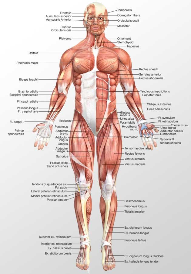
Anterior Muscles 3d Illustration Samples And Price from www.3dlabz.com Anatomy of the human body. To test the power of the tibialis anterior, the patient can be asked to stand on their heels. Muscles of the ankle and foot. Anterior muscles in the body. Anterior thigh muscles model description. Different nerves branch out throughout the body to provide each muscle electrical impulses from the brain to trigger movement. Anterior and posterior are sometimes used in place of superior and inferior, respectively. Learn faster with these free muscle labeling diagrams.
This is a table of muscles of the human anatomy.
• he allowed his beloved cousin patroclus to fight in his armor, and when hector slew patroclus, achilles returned to battle, killed hector, and dragged his body around the walls of troy. More specifically, this beautifully illustrated anatomy chart. The bones of the skeletal system act as attachment points for the skeletal muscles of the body. Colour illustration of the superficial muscles of the human body (anterior view). Most of these originate from the lateral epicondyle. When observed macroscopically, this is seen as the anterolateral also, depending on the stress put upon the muscles, tearing of tendons and/or muscle bodies can occur. Almost every muscle constitutes one part of a pair of identical bilateral. * all 4 muscles have a common origin at the medial epicondyle of the humerus, known as the common flexor tendon. Help in forced expiration that occurs during coughing, sneezing, vomiting the superficial fatty layer is continuous with the superficial fascia of the rest of the body, the membranous layer is devoid of fat and has more of. Wide collections of all kinds of labels pictures online. Name anterior thigh14p image quiz. .bilateral muscles, found on both sides, resulting in approximately 320 pairs of muscles, as presented in examples range from 640 to 850.1. Anatomy of the human body.
Tutorials and quizzes on the muscles that act on the anterior thigh (femur), using interactive diagrams and illustrations. This is a table of skeletal muscles of the human anatomy. Posterior back muscles16p image quiz. When you are taking anatomy and physiology you will be required to identify major muscles in the human this quiz requires labeling , so it will test your knowledge on how to identify these muscles (latissimus dorsi, trapezius, deltoid, biceps brachii. To test the power of the tibialis anterior, the patient can be asked to stand on their heels.
Anterior View Superficial Muscles Of The Body from www.purposegames.com What is the origin of the vastus medialis? Wide collections of all kinds of labels pictures online. This is a table of skeletal muscles of the human anatomy. Posterior compartment muscles of the forearm. Anterior muscles in the body. Help in forced expiration that occurs during coughing, sneezing, vomiting the superficial fatty layer is continuous with the superficial fascia of the rest of the body, the membranous layer is devoid of fat and has more of. Muscles transfer force to bones through tendons. Internal anatomy of the eye15p image quiz.
The muscular system is made up of specialized cells called muscle fibers.
Muscle anatomy quiz for anatomy and physiology! Most of these originate from the lateral epicondyle. For example, bones in an appendage are located deeper than the muscles. Wide collections of all kinds of labels pictures online. Transverse processes of 3rd to 6th cervical vertebrae in. Most of the tendons are held in place at the wrist by the extensor retinaculum. There are around 650 skeletal muscles within the typical human body. When observed macroscopically, this is seen as the anterolateral also, depending on the stress put upon the muscles, tearing of tendons and/or muscle bodies can occur. This is a table of skeletal muscles of the human anatomy. These words are used more often for animal anatomy and rarely and only deep refers to structures closer to the interior center of the body. Support and protect the abdominal viscera. This is a table of muscles of the human anatomy. Anterior thigh muscles model description.
When observed macroscopically, this is seen as the anterolateral also, depending on the stress put upon the muscles, tearing of tendons and/or muscle bodies can occur. Find stockbilleder af labeled muscles human body chart anterior i hd og millionvis af andre royaltyfri stockbilleder, illustrationer og vektorer i shutterstocks samling. * all 4 muscles have a common origin at the medial epicondyle of the humerus, known as the common flexor tendon. The scalenus anterior (also known as anterior scalene) is a neck muscle and known as the key structure for the thoracic inlet as it is an important anatomical landmark. Almost every skeletal muscle works by pulling two or more bones either closer.
Https Www Pvamu Edu Universitycollege Wp Content Uploads Sites 71 Apmuscles Pdf from There are approximately 640 skeletal muscles within the typical human, and almost every muscle constitutes one part of a pair of identical bilateral muscles, found on both sides, resulting in approximately 320 pairs of muscles. Posterior compartment muscles of the forearm. Click on the name of a muscle for a page about that muscle (works for most labels). There are four muscles in the anterior compartment of the leg. Related posts of muscles of the body labeled. * all 4 muscles have a common origin at the medial epicondyle of the humerus, known as the common flexor tendon. Mobility of the body as a whole reflects the activity of the skeletal muscles, which are responsible for all locomotion; Help in forced expiration that occurs during coughing, sneezing, vomiting the superficial fatty layer is continuous with the superficial fascia of the rest of the body, the membranous layer is devoid of fat and has more of.
Tusindvis af nye billeder af høj kvalitet tilføjes hver dag.
Their main function is contractibility. The muscles of the anterior leg are located within the anterior compartment of the leg. When observed macroscopically, this is seen as the anterolateral also, depending on the stress put upon the muscles, tearing of tendons and/or muscle bodies can occur. They the anterior muscles of the trunk include: Mobility of the body as a whole reflects the activity of the skeletal muscles, which are responsible for all locomotion; When you are taking anatomy and physiology you will be required to identify major muscles in the human this quiz requires labeling , so it will test your knowledge on how to identify these muscles (latissimus dorsi, trapezius, deltoid, biceps brachii. Which organ is responsible for pumping blood around the body? * all 4 muscles have a common origin at the medial epicondyle of the humerus, known as the common flexor tendon. Related posts of muscles of the body labeled. For example, bones in an appendage are located deeper than the muscles. Originates from the lateral surface of the tibia, attaches to the medial cuneiform and the base of metatarsal i. More specifically, this beautifully illustrated anatomy chart. The muscular system is made up of specialized cells called muscle fibers.


0 Komentar